Many of us may already come across the term MRI(Magnetic Resonance Imaging) in our day-to-day lives. I also had a bit of luck to take an MRI due to the ACL(Anterior cruciate ligament) injury two years back. The most painful thing was not the injury but the bill of MRI just for the knee. Now let’s look why it is so precious to loot your money and what is the technique behind MRI.

Magnetic Resonance Imaging (MRI) allows us to see inside the human body in detail. The first MRI scanner used to image the human body was built in New York in 1977. MRI does not involve radiation like an X-ray or CT scan so that it can be done frequently without any side effects.
Magnet in MRI
There is essentially a big magnet in the MRI scanner. Most of the MRI scanners used in hospitals and medical research clinics are 1.5 or 3 T (Tesla). It is around 60,000 times stronger than the earth’s magnetic field!.

The MRI scanner measures how much water is in different tissues of the body using a magnetic field and radio waves, maps the location of water, and uses this information to generate a detailed image. Luckily the human body has around 70% of water, and there are many signals to measure so that the image will be in detail and accurate. The hydrogen atoms are the part that makes water interesting for MRI, and what we use to measure the signal from the body when we do an MRI scan.
Hydrogen Atom and MRI
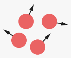
For hydrogen, we see it has a central nucleus containing a single positive charge, called a proton. Like the Earth spinning on its axis with a north and south magnetic pole, each spinning hydrogen proton is like a tiny magnet spinning on its axis. This spinning motion is known as precession. Billions of hydrogen protons in our bodies are all in random positions and spinning on their axes.
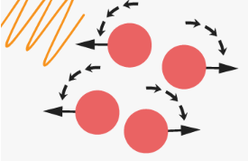
This randomness changes when we place a human body into a powerful magnetic field, like an MRI scanner. Just like a compass needle aligns with the Earth’s magnetic field. When randomly spinning hydrogen protons are placed in an MRI scanner, their axes realign them with the powerful magnetic field.

Some will align parallel, and some will align anti-parallel. The numbers will never be equal, but they all will still spin around their axis. Even though most magnetic moments cancel out, there will be a slight magnetic field in one direction due to the inequality in numbers. It is this small magnetic field that we can measure using MRI.
The powerful magnetic field from the magnet field not only affects the hydrogen proton’s alignment but also affects how fast these protons spin called precessional frequency. The stronger the magnetic field, the faster they spin. These two property of axis realignment and precessional frequency are the key ideas for MRI to measure the signals from hydrogen molecules.
Detecting the Magnetic Field
To distinguish the small magnetic field. We use something called a radio frequency (RF) pulse to disturb or flip all the protons, at the same time, out of alignment from the scanner’s magnetic field.
The frequency of the RF pulse must be the same as the frequency of the spinning hydrogen protons so that resonance occurs. Resonance enables the protons to absorb enough energy from the RF pulse to rotate their axes away from the strong field so that the MRI scanner can measure it. If we think again about our compass in the Earth’s magnetic field pointing toward the north pole, we can make the needle rotate to point east if we place a small bar magnet next to the compass. This is similar to the way the protons behave when we turn on the RF pulse.
Imaging From Spinning Protons
When the RF pulse is turned off, the protons flip back and realign along the main magnetic field. If we think of our compass again, when we move our small bar magnet away, the needle will rotate from east to north and align with the Earth’s magnetic field once more.

As the protons flip back and realign with the main strong magnetic field, they give off energy. Different tissues in the body give off different amounts of energy. To measure this emitted energy, we need a coil that we see, which is placed around the body part that we are imaging that detects the released energy as an electrical current. This electric current is converted into images via a computer using Fourier transformation.
I have now described how we use MRI to generate and measure signals from Hydrogen molecules in the body. But as well as providing images of the inside of our bodies, MRI can also be used to answer many different questions about the makeup of the brain and how it functions. From the MRI images, we can investigate the structure and chemical composition, how our mind’s network is connected, and how different regions of the brain communicate with each other.

Excellent !!
LikeLike